Radiology
24-07-2023
Publication date: 29-01-2024
Updated on: 29-01-2024
Topic: Radiology
Estimated reading time: 1 min
Article Author
Miriam Manfrini
Medical Editor
Pietro Panizza
Editor and Translator
Viktoryia LuhakovaMammography is the key exam for the diagnosis of breast cancer: the cancer with the highest estimated incidence in the global population (55,700 new diagnoses in Italy alone in 2022, which is 0.5% higher than in 2020).
However, according to the American Cancer Society, the 5-year survival from localized, noninvasive stage diagnosis is 99%, which proves the integral role of periodic breast screening and checkups, performed with modern technologies.
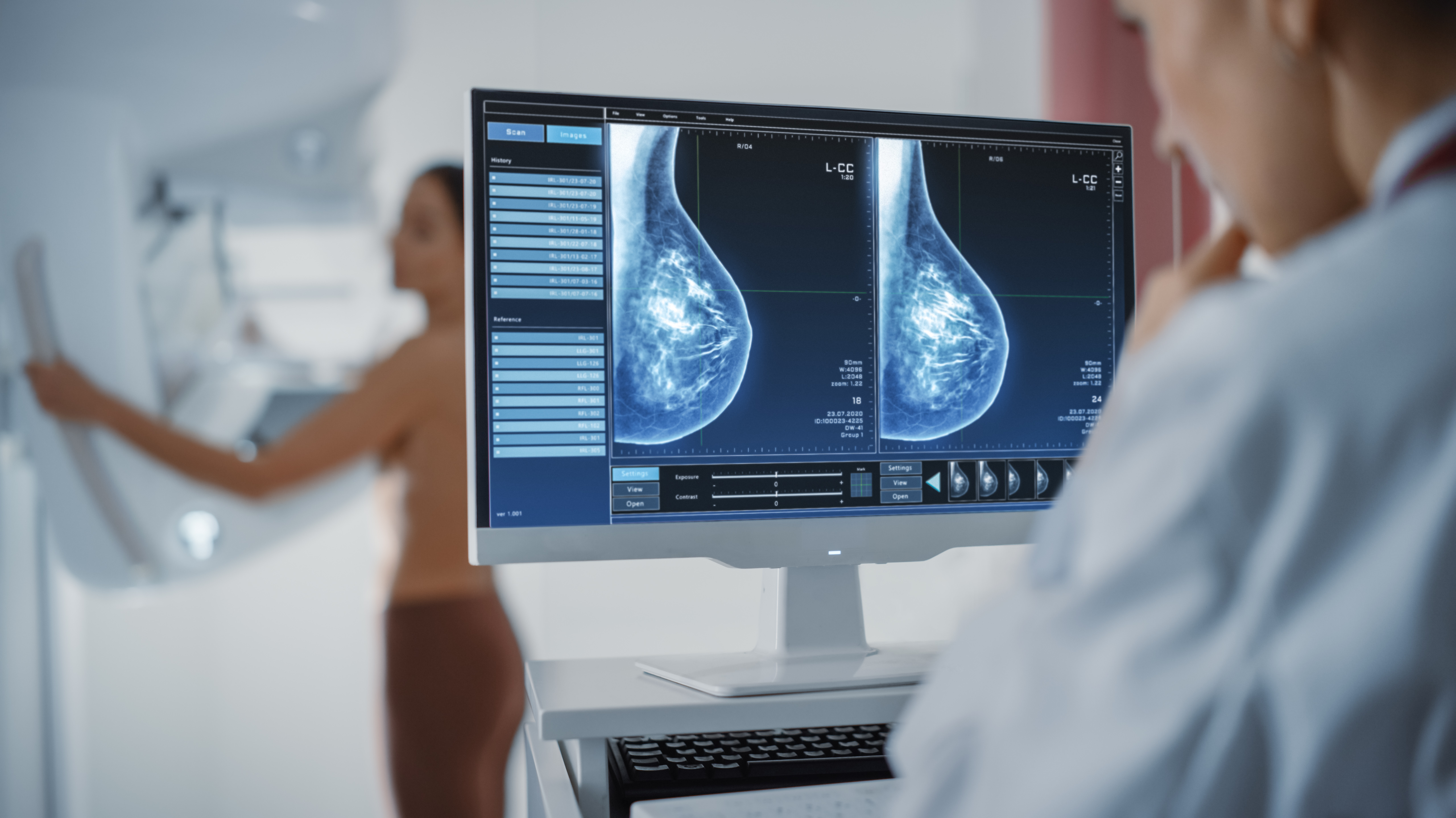
Dr. Pietro Panizza, a breast radiologist at Casa di Cura La Madonnina and Chief of the Breast Imaging Department at IRCCS Ospedale San Raffaele, explains what is the 3D-like mammography or tomosynthesis, which is already available at Casa di Cura La Madonnina.
"Tomosynthesis or Digital Breast Tomosynthesis (DBT)," the Doctor explains, "is an evolution of mammography with 2D technology. The tomosynthesis apparatus is identical to the 2D mammographer and similarly involves compression of the breast; however, thanks to an oscillating movement of the X-ray tube (which produces X-rays), tomosynthesis allows images to be acquired from different angles."
In this way, 3D-like breast images with volumetric acquisition and processing of numerous thin organ sections are obtained.
The advantages of 3D-like mammography or tomosynthesis, compared with the traditional 2D technique, are the following:
In fact, some studies have shown:
The duration of the examination is the same as the one of 2D mammography.
No special preparation procedures are required, but applying creams and deodorants to the breast area should be avoided on the day of the examination.
Mammography with tomosynthesis does not have any particular risks other than exposure to a low dose of ionizing radiation. These doses are much lower than those established by ICRP (International Commission on Radiological Protection) and considered safe because the risk-benefit ratio clearly favors the benefit associated with early detection.
Casa di Cura La Madonnina has recently upgraded the breast diagnostic area by acquiring a mammographer with tomosynthesis, and an ultrasound scanner with dedicated probes for breast study.
"These technologies facilitate accurate and timely breast diagnoses. By investing in state-of-the-art equipment, the clinic initiates a process of modernizing diagnostic technologies, improving operational efficiency and the diagnostic process," the doctor specifies.
Regular checkups are crucial in the fight against breast cancer as they allow to detect it at an early stage. For this reason, it is reccomended:
In case of palpable changes, ultrasonography is the most appropriate examination. In case of doubtful/suspicious images or dense sense at mammography, the Radiology Physician decides whether to complement it with ultrasonography.
When it comes to the risk of developing breast cancer, it's important to acknowledge factors we have some control over and those that are beyond our influence.
Non-modifiable risk factors include:
Modifiable risk factors, on the other hand, include: