Orthopedics
30-05-2025
Publication date: 22-09-2022
Updated on: 16-05-2025
Topic: Orthopedics
Estimated reading time: 1 min
Article Author
Elena Buonanno
Editor and Translator
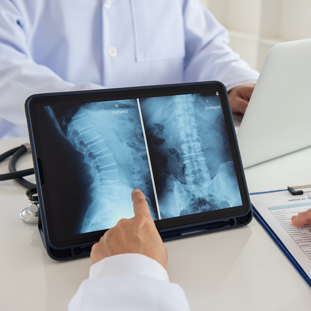
Viktoryia LuhakovaA complete x-ray of the spine is an examination that allows you to check the real condition and health of the spine and identify any problems in just 10 minutes. We spoke with Dr. Alberto Castoldi, Head of Radiology and Diagnostic Imaging unit at the Policlinico San Pietro and of the Radiology Service at the Smart Clinic Oriocenter, where this examination is performed.
A complete x-ray of the spine allows to obtain in a single image the 'photograph' of the entire vertebral column, from the cervical to the pelvis, and to be able to evaluate cervical, thoracic and lumbosacral vertebrae.
It is a safe and non-invasive test. The technologies available today allow it to be carried out in total safety: the doses of radiation currently used to perform the examination and the exposure time have been significantly reduced compared to past years.
The only contraindication, as indeed for other traditional radiographic investigations, is pregnancy.
Complete spine radiography is a safe and reliable examination that allows you to detect the presence of:
In addition to these deformations of the spinal column, the examination is able to identify anomalies in the load axes of the lower limbs and dysmetria of the pelvis due to scoliosis and / or different length of the lower limbs. Finally, it allows you to study the spine also on a postural level.
This examination usually lasts about 10 minutes and is carried out:
Once the x-ray is finished you can immediately return to everyday life.
There are no particular preparation rules. However, it is advisable to wear comfortable clothes and avoid wearing metal objects that should be removed during execution as they could interfere in obtaining the x-ray.
This radiograph can be requested by physiatrists, orthopedists and pediatricians with the aim of monitoring structural growth in particular in adolescents and in people suffering from arthrosis with hip or knee prostheses.