Cardiovascular diseases
07-11-2025
Publication date: 16-01-2023
Updated on: 16-05-2025
Topic: Cardiovascular diseases
Estimated reading time: 1 min
Article Author
Clelia Andolina
Medical Editor
Matteo Montorfano
Editor and Translator
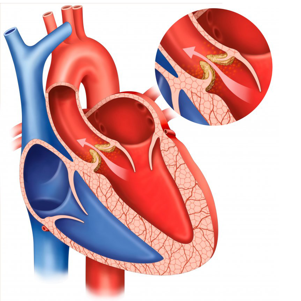
Viktoryia LuhakovaAortic valve stenosis is the most common heart valve disease in industrialized countries. It occurs when the aortic valve, which is located between the left ventricle (the most important part of the heart, which pumps blood throughout the body) and the aorta ("pipe" that carries blood to the entire body), malfunctions and blood struggles to pass through it, hindering the pumping function by the heart.
What kind of procedure is used when valve repair is needed and how is it performed? We discuss this with Dr. Matteo Montorfano, Chief of the Interventional Cardiology Unit at Ospedale San Raffaele.
Heart valve diseases, or valvulopathies, are diseases that affect the valves of the heart and their functioning. There are 4 valves:
These allow blood to pass between the various heart chambers and prevent reflux.
The most frequent valvulopathies are those of the left sections of the heart (mitral valve and aortic valve, i.e. the part of the heart that irrigates the whole body). Valvulopathies affecting the right sections of the heart (which instead carry blood to the lungs for oxygenation) are less common.
If the aortic valve is narrowed and blood struggles to pass through it, it is called valvular stenosis. On the other hand, when the valve does not close properly and blood flows back where it should not, we are dealing with valve insufficiency.
Aging is the leading cause of aortic valvulopathy in the stenotic sense: as time passes, the valve becomes very stiff, undergoes calcification, and this prevents the flaps that constitute it from opening properly, creating narrowing and thus difficulty in the passage of blood from the left ventricle to the aorta.
In the case of aortic insufficiency, valve degeneration causes a defect in the closure of the valve leaflets, which causes incontinence, resulting in the return/regurgitation of blood into the left ventricle, which is overworked.
Aortic stenosis has typical symptomatology: breathlessness, chest pain, and fainting during exertion. If left untreated, aortic stenosis, in the presence of symptoms, can lead to death from heart failure, a condition in which there is accumulation of water in the lungs and throughout the body.
Prompt treatment in the case of aortic stenosis is crucial: patients with symptomatic aortic stenosis, if left untreated, have a poor prognosis with a 50% mortality rate at 2 years. Therefore, it is important to treat the condition promptly.
In the presence of symptoms, such as breathlessness, chest pain on exertion, and fainting, the finding of a heart murmur on auscultation may make one suspect aortic valvulopathy.
The suspicion should then be confirmed with an echocardiogram, a noninvasive test that uses ultrasound and allows the specialist to see the morphology of the valve, the presence of calcifications, and confirm the presence of valve stenosis.
Traditional treatment consists of surgical aortic valve replacement: an "open-heart" procedure that requires general anesthesia, chest opening and the use of extracorporeal circulation (i.e., a machine that temporarily replaces the function of the heart and lungs). The surgeon removes the calcified aortic valve by replacing it with a prosthesis. Recovery time after such surgery is about 3-4 weeks.
Minimally invasive treatment of aortic stenosis has been developed in recent years: the Transcatheter Aortic Valve Implantation (TAVI) procedure. The procedure is performed in interventional cardiology laboratories, under local anesthesia and without the need to open the chest.
This is the new frontier of minimally invasive interventional cardiology as an alternative to classic cardiac surgery: avoiding sternum opening means reducing the incidence of perioperative complications and patient hospitalization, with extremely fast recovery times.
A transcatheter aortic valve prosthesis implantation or TAVI procedure can be used to treat aortic stenosis. In its complexity and delicacy, this procedure requires great expertise of the medical workers and of the entire team: through the puncture of an artery in the leg (which becomes the port of entry), the necessary instruments are inserted to bring the new miniature valve into the heart.
Before the surgery is performed, it is important for the patient to have a CT scan so as to visualize the aorta and femoral arteries, i.e. the "road" that will have to be taken to implant the new prosthesis, and to accurately choose the type and size of the prosthesis to be implanted.
The procedure in experienced hands lasts for about an hour. It is preformed under sedation, without general anesthesia and does not involve extracorporeal circulation.
The length of hospitalization for such surgeries is between 5 and 7 days.