الرفاهية
02-12-2025
تاريخ النشر : 14-11-2025
تحديث في : 14-11-2025
الموضوع:
الوقت المقدر للقراءة : 1 دقيقة
محرر طبي
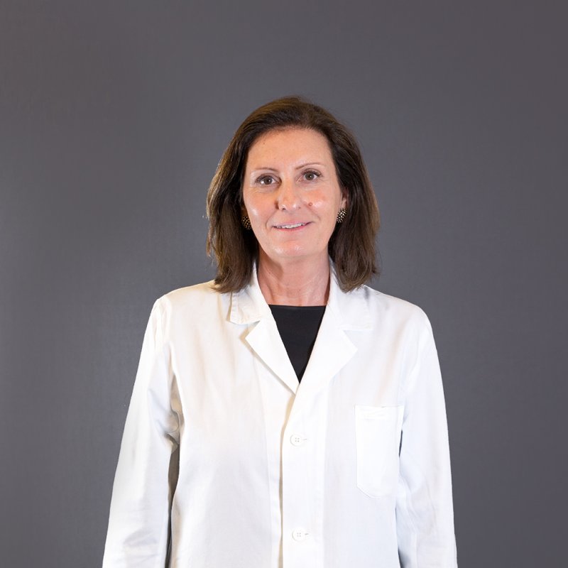
Maria Teresa Bonfanti
محرر ومترجم
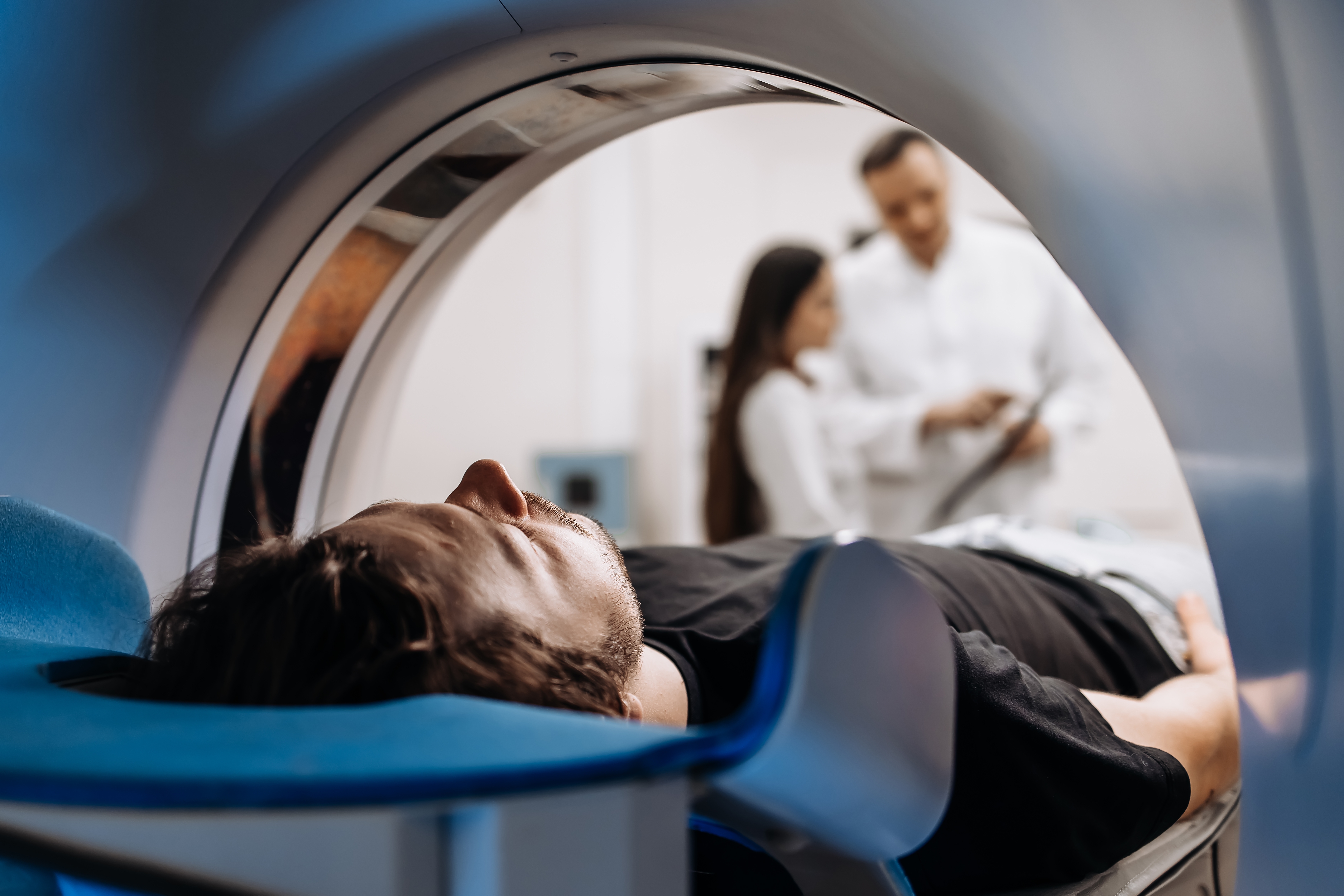
Viktoryia LuhakovaCasa di Cura La Madonnina takes an important step toward innovation with the introduction of a new, next-generation Magnetic Resonance Imaging system, combining modernity, speed, and safety. The upgrade significantly enhances clinical practice, patient comfort, and the operational efficiency of the diagnostic imaging department.
We explore the features of this device with Dr. Maria Teresa Bonfanti, Head of the Radiology Department at Casa di Cura La Madonnina.
The new MRI system at Casa di Cura La Madonnina uses artificial intelligence (AI) algorithms for image reconstruction.
“Thanks to a deep learning process based on the analysis of millions of anatomical cases, the system recognizes the normal appearance of the human body and integrates the patient’s images with reference models.
This technology, which enables the computer to ‘learn’ and identify complex patterns within images, allows us to obtain extremely high-quality scans in significantly shorter times,” explains Dr. Bonfanti.
The operator also receives suggestions on the most appropriate protocols, simplifying even the most complex examinations and improving diagnostic quality, including in technically challenging patients.
This imaging device is designed to reliably evaluate all major body regions:
All with image resolution improved by up to 65% and exam times up to three times faster compared to traditional technologies.
Its fully digital system minimizes background noise, while a 4K monitor provides exceptionally detailed visualization, enhancing diagnostic accuracy and clinical decision-making.
New 3D sequences also allow:
The technological advancements of this new MRI system transform diagnostic and therapeutic workflows across various clinical areas, including the prostate, heart, breast, and nervous system.
Modern equipment no longer requires the endorectal coil used in older scanners. The resulting images are equal or superior in quality.
The new MRI enables a complete 360° evaluation of the heart, assessing contractility, wall thickness, valvular function, and blood flow both at rest and under pharmacological stress.
Advanced technologies allow cardiac MRI to be performed with free breathing or short breath-holds, still maintaining excellent image quality. AI also accelerates processing and automatically identifies cardiac phases, making the examination faster and more efficient.
This system produces extremely detailed images of breast tissue and breast implants, even without contrast agent.
With AI software and algorithms, the scanner:
The new MRI can examine multiple components of the nervous system (brain, orbits, temporal bone, spinal cord) within a single integrated session, including evaluation of cerebral arteries crucial for preventing cerebrovascular disease.
State-of-the-art software and AI algorithms allow optimal assessment of common neurological disorders (degenerative, inflammatory, oncologic), as well as complex conditions such as Alzheimer’s disease, vasculitis, and epilepsy. They improve therapeutic planning and streamline workflow.
For the first time, Casa di Cura La Madonnina offers Whole-Body MRI, allowing full-body analysis in a single session. In under 60 minutes, the exam assesses every body region from brain to legs.
Thanks to high-definition whole-body imaging, this exam enables:
The exam’s speed and absence of ionizing radiation provide major advantages in terms of patient safety and comfort.
Whole-Body MRI raises the diagnostic standard, confirming Casa di Cura La Madonnina’s commitment to offering the most advanced technologies for patient care.
This new scanner integrates solutions that combine comfort, safety, and ease of use:
The result is a system that delivers maximum diagnostic quality while prioritizing patient experience, adapting to all ages, body types, and physical conditions.
The new MRI has also been designed with children in mind: